Magnetic Resonance Imaging (MRI) is a household name in healthcare, with an estimated 100-150 million Magnetic Resonance Imaging (MRI) scans performed globally each year to diagnose everything from brain tumors to torn ligaments. Its power is undisputed, but its process has always been slow, complex, and expensive. At Orbem, we saw an opportunity to change that. We are on a mission to industrialize MRI, transforming it into a tool that is fast, automated, and accessible to everyone, starting with the food industry.
To achieve this, we had to completely rethink how MRI works.
The Challenge: A Race Against the Clock
Taking poultry as an example, let’s look at how we adapt MRI for the purpose of sexing unhatched chicken eggs. The global poultry industry incubates approximately 100 billion eggs each year. If we used traditional medical MRI methods to look inside them, it would take us roughly 3.8 million years. A typical hospital MRI scan requires a patient to be manually positioned on a table, moved into the scanner, and then wait anywhere from 15 to 90 minutes for the scan to complete. Every step demands human interaction.
In our early days, we followed this manual process, scanning about one egg per minute. Today, our automated Genus Focus system scans an egg in less than one second. How did we achieve this leap in performance?
The Solution: A Symphony of Automation
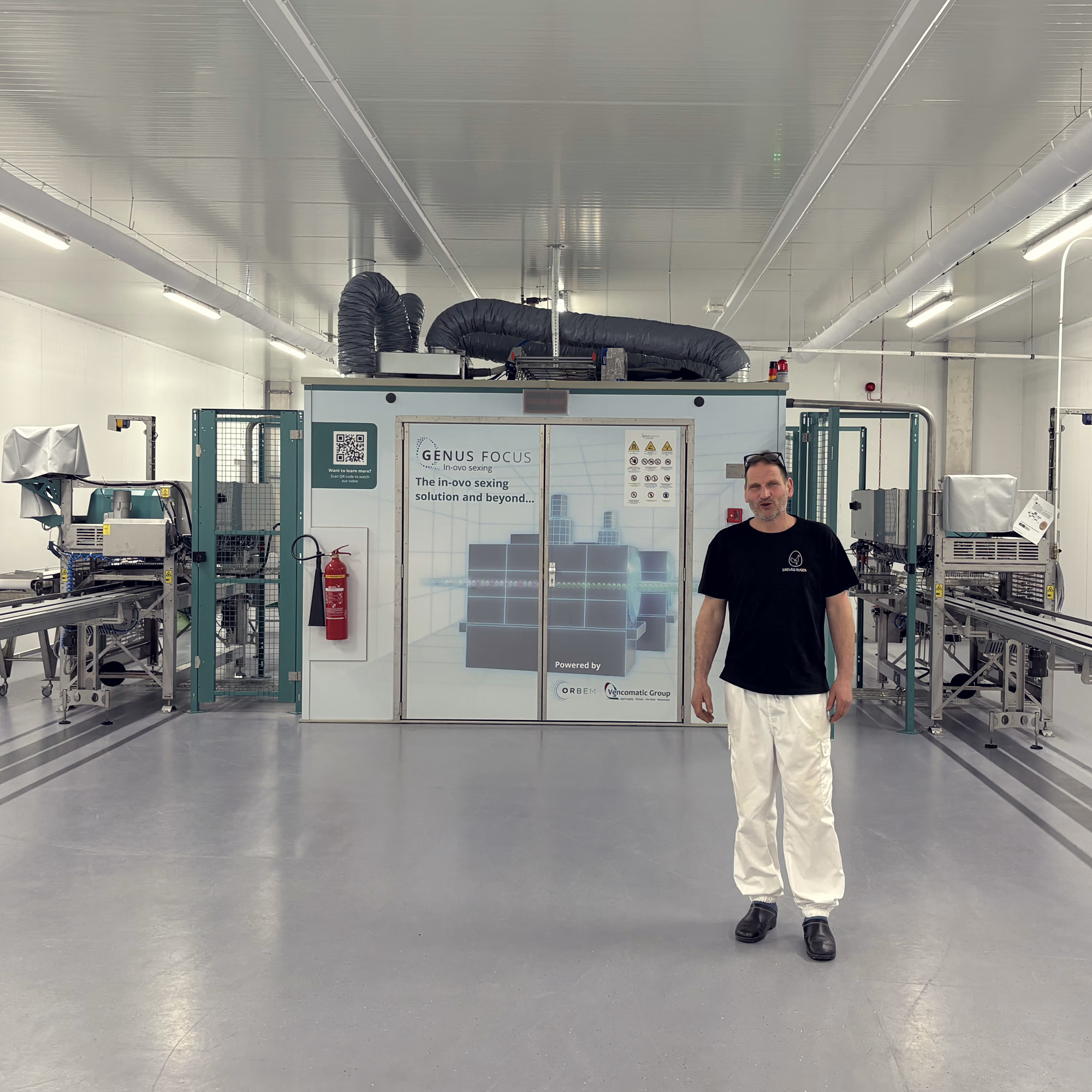
In close partnership with automation expert the Vencomatic Group, we built a fully automated system that eliminates the need for human interaction and seamlessly integrates in the hatchery process. This process has four key elements:
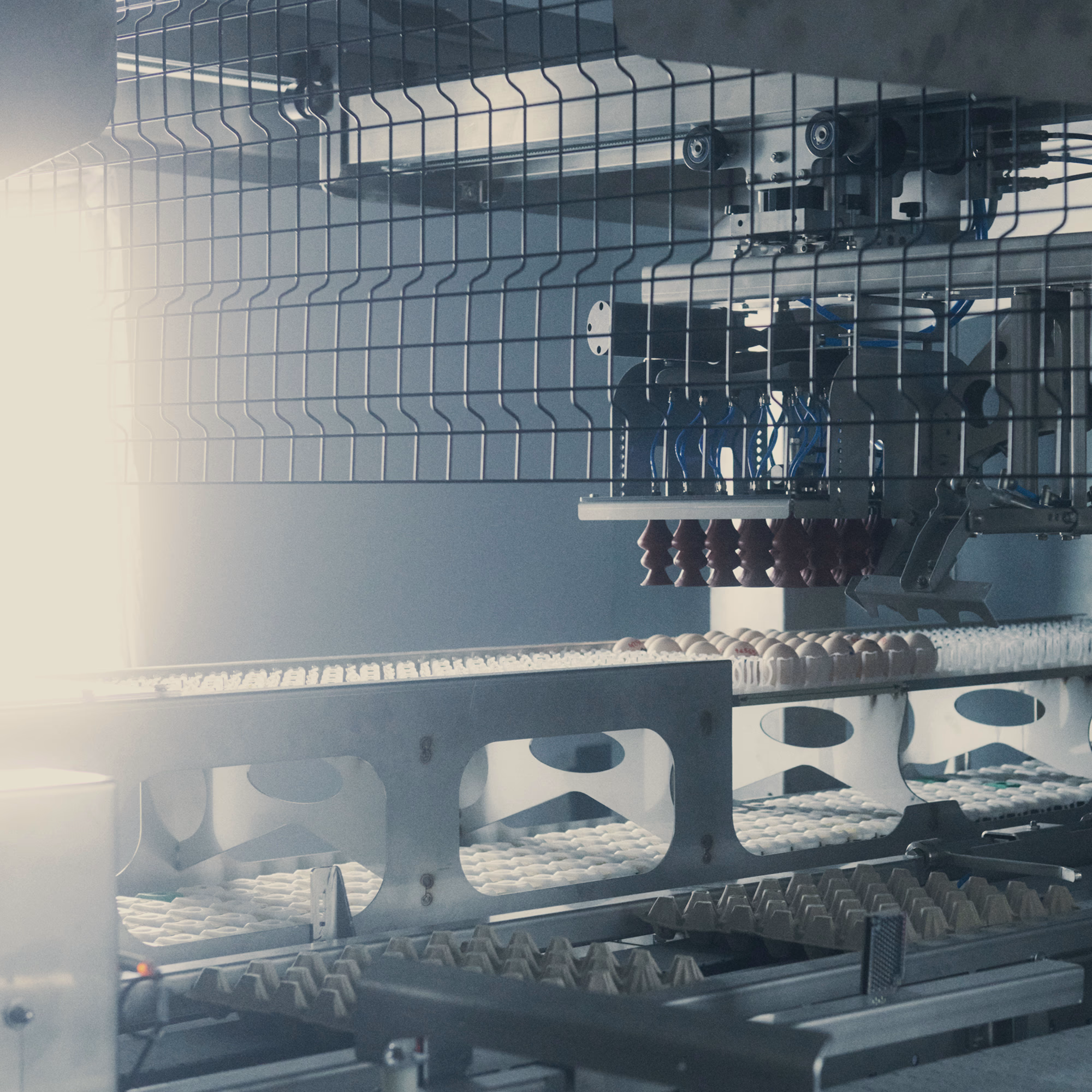
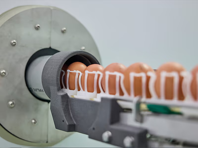
1. Automated Egg Handling
The process begins with a robotic pick-and-place system that gently lifts eggs from setter trays using silicone suction cups. Once a tray is empty, the system automatically proceeds it to the empty tray stacker and moves to the next, ensuring a continuous supply.
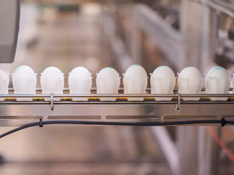
2. The Egg Chain
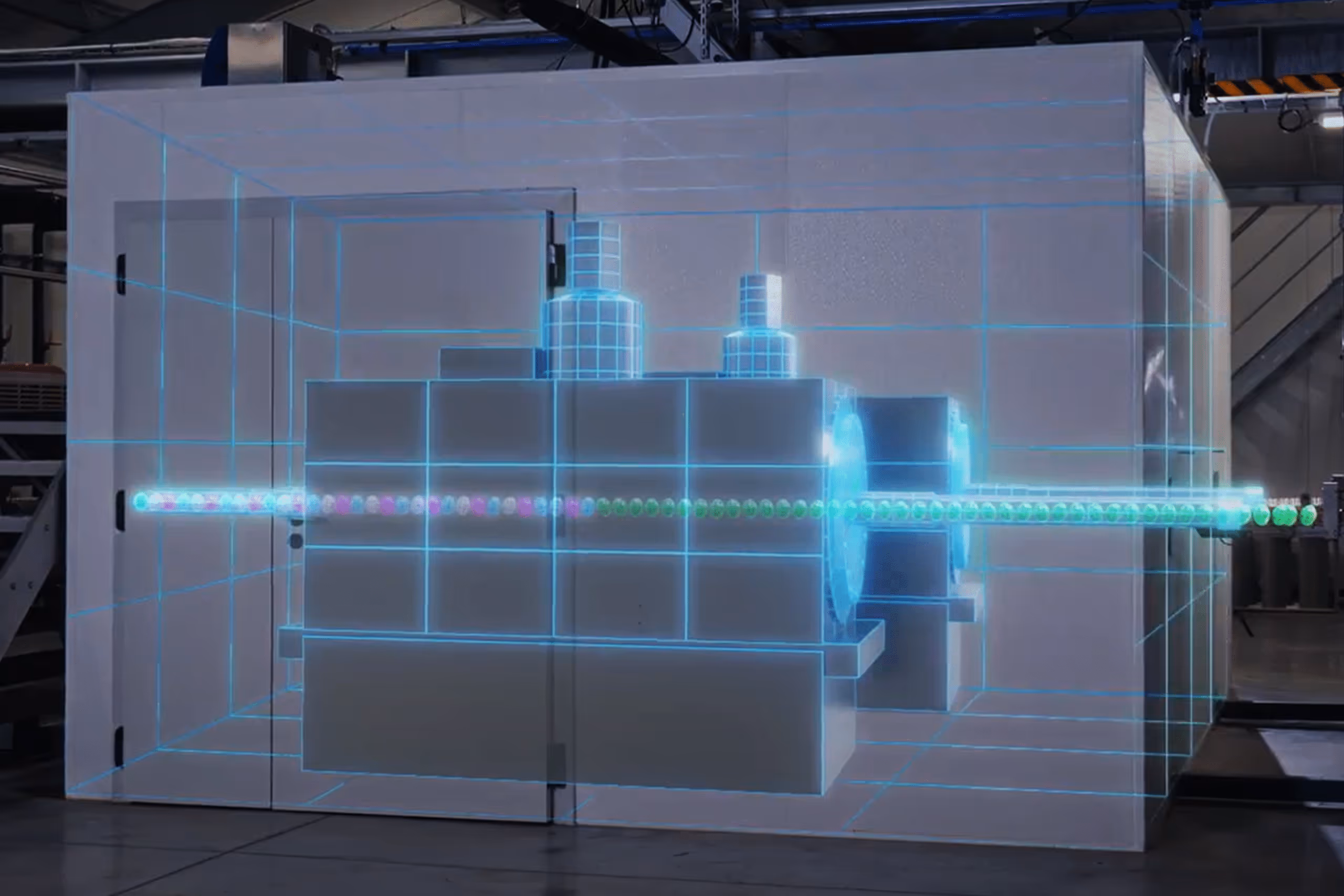
Instead of a slow patient table, we designed an “egg chain” that holds eggs in single file. This chain moves continuously through the center of the MRI scanner, with each egg advancing to the scanning position in just a fraction of a second. The scanner is almost always in operation, maximizing throughput.
3. Intelligent Scan Automation
The entire scanning process is a seamless digital conversation. The egg chain controller signals the MRI scanner to begin a scan the moment an egg is in position. Once finished, the scanner signals the chain to advance to the next egg. Simultaneously, the image data from the previous egg is sent to a server for AI classification, ensuring no time is lost.
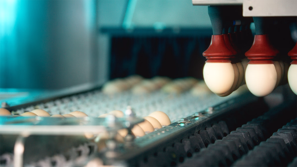
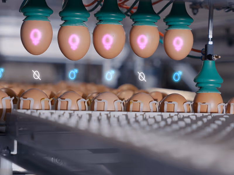
4. Precision sorting
On the outbound side, another robotic system sorts the eggs based on the AI’s classification results—accurately separating them into male, female, or unfertilized categories.
Overcoming the Challenges of Industrial MRI
Automating MRI on this scale required solving complex challenges:
Flawless data tracking
Our system manages multiple asynchronous processes—like egg movement, scanning, and AI classification—and must perfectly match every classification result to the correct physical egg on the chain. Mistakes here are not an option.
MRI-Safe Design
The powerful magnetic fields in an MRI scanner required us to construct the entire egg chain from non-metallic materials, like specialized plastics and rubber, to ensure safety and prevent image interference. For the same reason, our magnet room is locked at all times, to anyone except for MRI engineers. Because we have developed a reliable industrial version of MRI, our magnet rooms never need access outside of the yearly preventative maintenance by a trained MRI engineer.
The Challenge of Speed vs. Quality
Finally, our system must meet the immense throughput demands of modern hatcheries, where thousands of eggs require analysis daily. In imaging, there is a fundamental trade-off: accelerating an MRI scan typically reduces image quality. However, in our process, we cannot compromise on accuracy. This is where our deep expertise shines. Our team of MRI and AI scientists implements state-of-the-art acquisition and reconstruction techniques that overcome this challenge, ensuring maximum throughput without any expense to accuracy.
We are constantly investigating new methods to push our processing speeds even further, redefining what is possible in industrial imaging.
The Future of Food, Powered by MRI
By mastering the automation of MRI, Orbem is doing more than just looking inside eggs. We are building a platform with the potential to significantly reduce waste and improve quality across the entire food industry. Viewing MRI not just as a healthcare tool but as a powerful industrial solution allows us to process food at a scale that was previously impossible, paving the way for a more efficient and sustainable global food system.
What is MRI?
- Magnetic Resonance Imaging (MRI) is an imaging modality that uses time-varying magnetic fields to produce images from within an object.
- The MRI scanner normally consists of a large magnet, giving the MRI scanner its distinctive cylindrical shape. Typically, this magnet produces magnetic fields up to 7 Tesla (the magnetic field of the Earth is 0.0005 Tesla). The hollow inside the magnet is known as the bore. Inserted within the bore are additional coils used to generate images: Gradient coils and a radiofrequency coil.
- Gradient coils produce linearly varying magnetic fields in the x, y and z directions. These are responsible for image creation. The radiofrequency coil behaves like a radio antenna, in that it transmits and receives electromagnetic waves to and from the object within the scanner.
- Unlike other imaging techniques, which detect object density (which is why X-ray, for example, is so often used in orthopedics or dentistry), MRI detects the electromagnetic signal from Hydrogen atoms in water (conveniently the human body is ~60% water).
- MRI is also a very versatile modality because of how the many ways that the magnetic fields of an MRI system can be played out will lead to very different images. We can see the organs inside the body and we can also, for example, distinguish between grey and white matter in the brain; healthy and cancerous tissue – and so much more.
About the Author

Benjamin Knowles, Staff MRI Integration Engineer
Benjamin specializes in automation and scalability in industrial MRI. Before joining Orbem, Benjamin did research at the German Cancer Research Center (DKFZ), at King’s College London, and at Harvard Medical School.